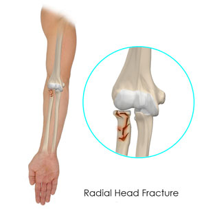
Elbow Anatomy
The arm in the human body is made up of three bones that join to form a hinge joint called the elbow. The upper arm bone or humerus connects from the shoulder to the elbow to form the top of the hinge joint. The lower arm or forearm consists of two bones, the radius, and the ulna. These bones connect the wrist to the elbow forming the bottom portion of the hinge joint.
The elbow joint is essential for the movement of your arms and to perform daily activities. The head of the radius bone is cup-shaped and corresponds to the spherical surface of the humerus. The injury in the head of the radius causes impairment in the function of the elbow.
What are Radial Head Fractures of the Elbow?
Radial head fractures are very common and occur in almost 20% of acute elbow injuries. Elbow dislocations are generally associated with radial head fractures. Radial head fractures are more common in women than in men and occur more frequently in the age group of 30 to 40 years.
What are the Causes of Radial Head Fractures?
The most common cause of a radial head fracture is breaking a fall with an outstretched arm. Radial head fractures can also occur due to a direct impact on the elbow, a twisting injury, sprain, dislocation or strain.
Symptoms of Radial Head Fractures
The symptoms of a radial head fracture include severe pain, swelling in the elbow, difficulty in moving the arm and visible deformity, which can indicate dislocation, bruising and stiffness.
How are Radial Head Fractures Diagnosed?
Your doctor may recommend an X-ray to confirm the fracture and assess displacement of the bone. Sometimes, your doctor might suggest a CT scan to obtain further details of the fracture, especially the joint surfaces.
What are the Treatment Options for Radial Head Fractures?
The treatment of a fracture depends on the type of fracture.
- Type 1 fractures are usually very small. The bone appears cracked but remains fitted together. The doctor may use a splint (casting) to fix the bone. You may have to wear a sling for a few days. If the crack becomes intense or the fracture gets deep, then your doctor may suggest surgical treatment.
- Type 2 fractures are characterized by displacement of bones and breaking of bones in large pieces and can be treated by surgery. During surgery, your doctor will correct the soft-tissue injuries and insert screws and plates to hold the displaced bones together firmly. Small pieces of bone may be removed if it prevents the normal movement of the elbow.
- Type 3 fractures are characterized by multiple broken pieces of bone. Surgery is considered the compulsory to either fix or remove the broken pieces of bone, sometimes including the radial head. An artificial radius head may be placed to improve the function of the elbow.


